Specimen Model Service Manufacturer
Manufacturing, production and sales

13506223680
Specimen Model Service Manufacturer
Manufacturing, production and sales
13506223680

Zhangjiagang Bailing Specimen Model Co., Ltd.
Contact:Mr Zhang
Eelephone:13506223680
Eelephone:18962473680
E-mail:cnbbmx@163.com
605340177@qq.com
Website:www.cnbbmx.com
Address:Bridge No. 4, Leyu Town, Zhangjiagang City, Suzhou City
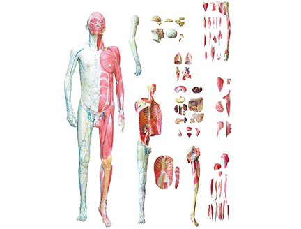
mannequin muscle anatomy
The muscles of the human body can be divided into trunk muscles, upper extremity muscles, lower extremity muscles and head and neck muscles according to their parts.
trunk muscles
The trunk muscles include the dorsal, pectoral, diaphragm, abdominal, and perineal muscles.
The back muscles are divided into superficial and deep layers. The superficial dorsal muscles include the trapezius, latissimus dorsi, levator scapulae, and rhomboids. The deep dorsal muscles are divided into the longus dorsi and the brevis. The longus dorsi muscles include the erector spinae and the splenius. The brevis muscle includes the transverse process spinous muscle, the interspinous muscle and the intertransverse process muscle (including the semispinalis, rotator, and multifidus three parts).
The pectoralis muscle is divided into the upper extremity pectoralis muscle and the pectoralis propria muscle. The upper thoracic muscles include the pectoralis major, pectoralis minor, and serratus anterior. The pectoralis propria muscles include the external intercostal muscles, the internal intercostal muscles, and the transverse pectoralis.
The abdominal muscles include the rectus abdominis, external oblique, internal oblique, and transverse abdominis of the anterior abdominal wall and the quadratus lumborum of the posterior abdominal wall.
upper extremity muscles
Upper extremity muscles include shoulder girdle, upper arm, forearm, and hand muscles.
The shoulder girdle muscles originate from the clavicle and scapula and insert into the humerus. Includes deltoid, supraspinatus, infraspinatus, teres minor, subscapularis, and teres major. Among them, the tendons of the supraspinatus, infraspinatus, teres minor and subscapularis together form a structure called "tendon sleeve" (also known as "rotator cuff"), which strengthens and protects the shoulder joint
The muscles of the upper arm wrap around the humerus and are divided into two groups: anterior and posterior. The anterior group (flexor group) includes the biceps, coracobrachialis, and brachialis. The posterior group (extensor group) includes the triceps and elbows.
Forearm muscles have a high degree of differentiation, mostly long muscles with long tendons. They are divided into two groups: front and back, and each group is divided into superficial and deep layers. The front muscles are located on the front and inside of the forearm, and the rear muscles are located on the back and outside of the forearm. The superficial layers of the anterior muscles are mainly brachioradialis, pronator teres, flexor carpi radialis, and flexor carpi ulnaris. The superficial layers of the posterior group of muscles mainly include extensor carpi radialis longus, extensor carpi radialis brevis, and extensor carpi ulnaris.
lower extremity muscles
Lower extremity muscles include the pelvic girdle, thigh, calf, and foot muscles.
The pelvic girdle muscles are divided into anterior and posterior groups. The anterior cluster originates from the inner surface of the pelvis, and the posterior cluster originates from the outer surface of the pelvis. The anterior group (medial group) has the iliopsoas and piriformis. The posterior (lateral) group includes the gluteus maximus, gluteus medius, and gluteus minimus.
The thigh muscles can be divided into anterolateral, posterior and medial groups. The anterolateral group includes the quadriceps, sartorius, and tensor fascia lata. The posterior group includes the biceps femoris, semitendinosus, and semimembranosus. The biceps femoris, semitendinosus, and semimembranosus muscles together are called the hamstrings or hamstrings. The medial group includes the pubic muscle, the adductor longus, the adductor brevis, the adductor magnus, and the gracilis.
The calf muscles are divided into anterior, posterior and lateral groups. The anterior group includes the tibialis anterior and the extensor digitorum longus. The posterior group includes the triceps calf, flexor digitorum longus, flexor hamstring longus, and tibialis posterior. The lateral group has the peroneus longus and peroneus brevis.
neck muscles
In the head and neck muscles, the head muscles can be divided into expressive muscles and masticatory muscles; the cervical muscles are divided into three groups: superficial, medium and deep. The superficial cervical muscles include the platysma and sternocleidomastoid.
sternocleidomastoid muscle
Location: Deep layer of the platysma, on both sides of the neck.
Origin: manubrium and sternal end of clavicle.
Insertion: mastoid of temporal bone.
Function: During lower fixation, one side is contracted to bend the head and neck to the same side and turn to the opposite side; when both sides are contracted, the line of action of the resultant muscle force is behind the frontal axis of the atlanto-occipital joint to extend the head, and the line of action of the resultant muscle force is on the atlanto-occipital joint. The front of the joint frontal axis flexes the head. When the upper is fixed, lift the chest up to help inhale.
sternocleidomastoid muscle
Rhomboid muscle
Location: Deep trapezius muscle.
Origin: Spinous processes of the 6th and 7th cervical vertebrae and 1st to 4th thoracic vertebrae.
Insertion: medial border of scapula.
Function: Raise, retract and rotate the scapula when near immobilization. During distal fixation, the sides contract to extend the thoracic spine.
Rhomboid muscle
erector spinae
Location: On both sides of the spine, it is composed of three parts: the spinous muscle, the longissimus muscle, and the iliocostalis muscle.
Origin: Dorsal sacrum, posterior iliac crest, lumbar spinous processes, and thoracolumbar fascia.
Insertion: Spinous and transverse processes of cervical and thoracic vertebrae, mastoid process of temporal bone, and costal angle.
Function: During lower fixation, one side contracts to flex the spine to the same side; both sides contract to extend the head and spine. Tilt the pelvis anteriorly during the upper fixation.
erector spinae
latissimus dorsi
Location: subcutaneous on the back and posterolateral side of the chest.
Origin: 7th to 12th thoracic vertebrae and all lumbar vertebrae spinous processes, median sacral ridge, posterior iliac crest, and lateral surfaces of 10th to 12th ribs.
Insertion: Lesser tuberosity ridge of humerus.
Function: Extends, adducts and internally rotates the shoulder joint during near immobilization. When far immobilized, pull the trunk closer to the upper arms and assist the inhalation.
latissimus dorsi
deltoid
Location: Under the skin of the shoulder, in an inverted triangle shape.
Origin: lateral half of clavicle, acromion and spine of scapula.
Insertion: Deltoid tuberosity of body of humerus.
Function: When near fixation, the contraction of the anterior fibers causes the shoulder joint to flex, horizontally flex and internally rotate; the contraction of the middle fibers causes the shoulder joint to abduct; the posterior fiber contraction causes the shoulder joint to extend, horizontally extend and externally rotate; the overall contraction can make the shoulder joint Joint abduction.
deltoid
Anatomy of the Rotator Cuff and Teres Major:
supraspinatus
Location: In the supraspinatus fossa of the scapula.
Origin: supraspinatus fossa of scapula.
Insertion: Greater tuberosity of humerus.
Function: To abduct the shoulder joint during near immobilization.
infraspinatus
Location: In the infraspinatus fossa of the scapula.
Origin: Infraspinatus fossa of scapula.
Insertion: Greater tuberosity of humerus.
Function; external rotation, adduction and extension of the shoulder joint when near immobilization.
teres minor
Location: Below the infraspinatus muscle.
Origin: Back of the lateral border of the scapula.
Insertion: Greater tuberosity of humerus.
Function: External rotation, adduction and extension of the shoulder joint during near immobilization.
shoulder girdle
Rotator cuff and teres major
Subscapularis:
Location: In the subscapular fossa of the scapula.
Origin: Subscapular fossa.
Insertion: Lesser tuberosity of humerus.
Function: In close fixation, internal rotation and adduction of the shoulder joint.
Teres major:
Location: below the infraspinatus and teres minor.
Origin: On the back of the lower corner of the scapula.
Insertion: Lesser tuberosity ridge of humerus.
Function: When near immobilization, internal rotation, adduction and extension of shoulder joint.
shoulder girdle
Biceps:
Location: The superficial layer in front of the upper arm, with long and short ends.
Origin: The long head originates from the superior glenoid tubercle of the scapula, and the short head originates from the coracoid process of the scapula.
Insertion: radial tuberosity and forearm fascia.
Function: flexes the shoulder joint, flexes and externally rotates the elbow joint when near immobilization. For distal fixation, bring the upper arm closer to the forearm.
Biceps
Triceps:
Location: Behind the upper arm, there are three heads: the long head, the lateral head and the medial head.
Origin: The long head originates from the infraglenoid tubercle of the scapula, the lateral head originates from the lateral and superior radial groove behind the humeral body, and the medial head originates from the medial and inferior radial groove.
Insertion: Olecranon ulna.
Function: When near immobilization, the elbow joint can be extended, and the long head can also extend the shoulder joint. For distal fixation, extend the upper arm at the elbow joint.
elbow muscle:
Location: Located behind the elbow joint, triangular in shape.
Origin: From the lateral epicondyle of the humerus.
Insertion: Inserts on the upper part of the back of the ulna.
Function: Extend and strengthen the elbow joint when it is near immobilization. For distal fixation, extend the upper arm at the elbow joint.
triceps
Serratus Anterior:
Location: The superficial layer of the lateral side of the thorax.
Origin: The lateral surface of the upper 8th to 9th ribs.
Insertion: Medial border of scapula and anterior to lower corner.
Function: When near immobilization, the scapula is extended forward; the lower muscle fiber contraction can make the scapula descend and rotate upward. when fixed at a distance.
serratus anterior
pectoralis major:
Location: subcutaneous on the upper part of the chest.
Origin: the medial half of the clavicle, the front of the sternum, the 1st to 6th costal cartilages, and the upper part of the anterior wall of the rectus sheath.
Insertion: ridge of the greater tuberosity of the humerus.
Function: Flexion, horizontal flexion, adduction and internal rotation of the shoulder joint during near immobilization. When the distance is fixed, pull the trunk closer to the upper arms, and lift the ribs to help inhale.
pectoralis major
pectoralis minor:
Location: Deep pectoralis major.
Origin: Anterior to the 3rd to 5th ribs.
Insertion: coracoid process of scapula.
Function: When near immobilization, make the scapula forward, descend and rotate downward. During distal fixation, lift the ribs to assist inhalation.
pectoralis minor
Brachialis:
Location: Deep in the lower half of the biceps.
Origin: Anterior lower half of humerus.
Insertion: ulnar tuberosity and coronoid process.
Function: flexes the elbow joint when near immobilization. For distal fixation, bring the upper arm closer to the forearm.
coracobrachialis:
Location: Located on the inside of the upper half of the biceps, it is the long fusiform muscle.
Origin: From the coracoid process of the scapula.
Insertion: Inserts on the medial side of the middle of the humerus (corresponding to the deltoid tuberosity).
Function: In near immobilization, it flexes, adducts and externally rotates the shoulder joint.
Brachialis, coracobrachialis and origin and insertion point
Brachioradialis:
Origin: Above the lateral epicondyle of the humerus
Insertion: radial styloid process.
Function: When near immobilization, the elbow joint is flexed, and the forearm is internally or externally rotated and maintained in a neutral position.
Flexor carpi radialis:
Origin: Medial epicondyle of humerus and forearm fascia.
Insertion: base of second metacarpal bone.
Function: flexes the radiocarpal joint during near fixation, participates in hand joint abduction, assists elbow flexion and forearm internal rotation.
Flexor carpi ulnaris:
Origin: Medial epicondyle of humerus, fascia of forearm and olecranon of ulna.
Insertion: pisiform bone, base of second metacarpal bone.
Function: In near immobilization, it flexes the radiocarpal joint, participates in adduction of the radiocarpal joint and elbow flexion.
Anterior superficial muscles of forearm
Pronator teres:
Origin: Medial epicondyle of humerus and coronoid process of ulna.
Insertion: the middle of the lateral surface of the radius.
Function: When near immobilization, the forearm is internally rotated to assist elbow flexion.
pronator teres
extensor carpi ulnaris:
Origin: Lateral epicondyle of humerus, forearm fascia and elbow joint capsule.
Insertion: base of fifth metacarpal bone.
Function: During near fixation, the radiocarpal joint is extended and involved in the adduction of the hand joint.
extensor carpi radialis longus:
Origin: Lateral epicondyle of humerus.
Insertion: base of second metacarpal bone.
Function: When near immobilization, extend the hand joint, participate in the abduction of the radiocarpal joint and the extension of the elbow joint.
extensor carpi radialis brevis:
Origin: Lateral epicondyle of humerus.
Insertion: base of third metacarpal bone.
Function: Basically the same as the extensor carpi radialis longus.
The superficial muscles of the posterior group of the forearm and their origin and insertion point
iliopsoas:
Location: Both sides of the lumbar spine and in the iliac fossa, consisting of the psoas major and iliacus.
Origin: The psoas major originates from the lateral surface and transverse process of the 12th thoracic vertebra and the 1st to 5th lumbar vertebrae; the iliacus originates from the iliac fossa.
Insertion: Lesser trochanter of femur.
Function: Flexion and external rotation of the hip joint during near immobilization. During distal fixation, one side contracts to flex the spine ipsilaterally; both sides contract to flex the spine and tilt the pelvis forward.
iliopsoas muscle
piriformis muscle:
Location: Anterior to the sacrum, posterior wall of the small pelvis.
Origin: Anterior aspect of the 2nd to 5th sacral vertebrae.
Insertion: Greater trochanter of femur.
Function: To abduct and externally rotate the hip joint during near immobilization. During distal fixation, one side is contracted to turn the pelvis to the opposite side; both sides are contracted to tilt the pelvis posteriorly.
piriformis muscle
Glutes:
Location: posterolateral side of the pelvis, subcutaneously on the buttocks.
Origin: outside the iliac wing and on the back of the sacrum and coccyx.
Insertion: femorogluteal tuberosity and iliotibial band.
Function: When near immobilization, the hip joint can be extended and externally rotated; the upper muscle fibers can contract to abduct the hip joint, and the lower part can make the hip joint adduction. During distal fixation, one side is contracted to turn the pelvis to the opposite side; both sides are contracted to tilt the pelvis posteriorly.
gluteus maximus
Gluteus medius and minimus:
Location: Outside the iliac wing, the back of the gluteus medius lies deep in the gluteus maximus, and the gluteus minimus lies deep in the gluteus medius.
Origin: Outside the ilium wing.
Insertion: Greater trochanter of femur.
Function: Abducts the hip joint when near immobilization; flexes and internally rotates the hip joint anteriorly. The rear extends and externally rotates the hip. During distal fixation, the contraction of one side causes the pelvis to tilt to the same side; the contraction of the anterior muscle fibers on both sides causes the pelvis to tilt forward, and the contraction of the posterior muscle fibers causes the pelvis to tilt backward.
Gluteus medius and minimus
Quadriceps:
Location: The front of the thigh, with four heads.
Origin: The rectus femoris muscle originates from the anterior inferior iliac spine; the vastus medius originates from the anterior aspect of the femoral body; the vastus lateralis muscle originates from the lateral lip of the femoral thick line; the vastus medialis muscle originates from the medial lip of the femoral thick line.
Insertion: The four heads merge into a single tendon that wraps around the patella and forms the patellar ligament down to the tibial tuberosity.
Function: When near immobilization, the rectus femoris muscle can flex the hip joint, and the overall contraction can extend the knee joint. When the distance is fixed, the thigh is extended at the knee joint to maintain the upright posture of the human body.
quadriceps
Pubic muscle:
Location: The superficial layer of the upper inner thigh.
Origin: Superior pubic ramus.
Insertion: The upper part of the medial lip of the thick line of the femur.
Function: Adducts, externally rotates and flexes the hip joint during near immobilization. During distal fixation, the sides contract to tilt the pelvis forward.
Adductor long and short adductor
Location: The adductor longus is located medially to the pubic muscle. The adductor brevis lies deep within the pubic and long adductor muscles.
Origin: The adductor longus originates from the outside of the superior pubic ramus, and the adductor brevis originates from the outside of the inferior pubic ramus.
Insertion: The adductor longus inserts at the middle of the medial lip of the femoral thick line, and the short adductor muscle inserts on the upper femoral thick line.
Function: Adducts, externally rotates and flexes the hip joint during near immobilization. During distal fixation, the sides contract to tilt the pelvis forward.
pubic muscle
Adductor magnus:
Location: Deep inner thigh.
Origin: ischial tuberosity, ischial ramus, and inferior pubic ramus.
Insertion point: the upper 2/3 of the medial lip of the femoral thick line and the medial epicondyle of the femur.
Function: Adducts, extends and externally rotates the hip joint during near immobilization. During distal fixation, the sides contract to tilt the pelvis posteriorly.
adductor magnus
Semitendinosus, semimembranosus, and biceps femoris make up the hamstrings
Biceps femoris:
Location: The superficial layer of the posterolateral aspect of the thigh, with long and short heads.
Origin: The long head originates from the ischial tubercle, and the short head originates from the lower half of the lateral lip of the femoral thick line.
Insertion: fibular head.
Function: When near immobilization, it can flex and externally rotate the knee joint, and the long head can also extend the hip joint. During distal fixation, contract both sides to flex the thigh at the knee; when the calf is straight, the pelvis is tilted back.
biceps femoris
Semitendinosus and semimembranosus:
Location: Posterior medial thigh, semimembranosus in the deep layer of semitendinosus. The lower half of the semitendinosus is the tendon, and the upper half of the semimembranosus is the aponeurosis.
Origin: ischial tuberosity.
Insertion: The semitendinosus muscle inserts on the medial side of the upper end of the tibia, and the semimembranosus muscle inserts behind the medial condyle of the tibia.
Function: When near immobilization, it can flex and internally rotate the knee joint, and can also make the hip joint extend. In distal fixation, the same as the biceps femoris.
semitendinosus and semimembranosus
Tibialis anterior:
Location: The anterolateral superficial layer of the calf.
Origin: the upper 2/3 of the lateral side of the tibia.
Insertion: medial cuneiform and base of 1st metatarsal.
Function: When near immobilization, the ankle joint is extended (dorsiflexed) and inversion. During distal fixation, the calf is extended at the ankle joint to maintain the arch of the foot.
tibialis anterior
Triceps Calf:
Location: The back of the calf. Including the superficial gastrocnemius and the deep soleus.
Origin: The medial and lateral heads of the gastrocnemius originate from the medial and lateral epicondyles of the femur, respectively. The soleus muscle originates from the posterior upper part of the tibia and fibula
End point: with nodules.
Function: In near immobilization, the ankle joint is flexed (plantar flexion), and the gastrocnemius muscle can also flex the knee joint. During distal fixation, the calf can be flexed at the ankle joint to assist the knee joint extension and maintain the uprightness of the human body.
calf triceps
soleus muscle
Transversus abdominis:
Location: Deep layer of internal oblique muscle. Muscle fibers are distributed laterally.
Origin: Medial surface of ribs 7-12, lateral to thoracolumbar fascia, iliac crest, and inguinal ligament
Stop point: white line. Its aponeurosis participates in the formation of the posterior wall of the rectus sheath.
Function: maintain abdominal pressure.
transversus abdominis
Rectus abdominis:
Location: Both sides of the midline of the anterior abdominal wall.
Origin: The upper border of the pubis.
Insertion: xiphoid process of the sternum and anterior to the 5th to 7th costal cartilages.
Function: During upper fixation, the sides contract to make the pelvis tilt backwards. During lower fixation, one side is contracted to flex the spine to the same side;
Contract the sides to flex the spine. You can also lower your ribs to help exhale.
rectus abdominis
Internal oblique muscle:
Location: Deep layer of the external oblique muscle. Muscle fibers run obliquely from the back to the bottom to the front to the inside.
Origin: thoracolumbar fascia, iliac crest and lateral inguinal ligament.
Insertion point: the lower border of the 10th to 12th ribs and the linea alba, and its aponeurosis participates in the formation of the anterior and posterior walls of the rectus abdominis sheath.
Function: During upper fixation, the sides contract to make the pelvis tilt backwards. During lower fixation, one side is contracted to flex and rotate the spine to the same side;
The sides contract to flex the spine.
internal oblique muscle
External oblique muscle:
Location: Anterior abdomen

Eelephone:13506223680 landline:0512-58961302
fax:0512-58961302 Website:www.cnbbmx.com
Address:Bridge No. 4, Leyu Town, Zhangjiagang City, Suzhou City
|
| |
cell phone station | WeChat public account |