Specimen Model Service Manufacturer
Manufacturing, production and sales

13506223680
Specimen Model Service Manufacturer
Manufacturing, production and sales
13506223680

Zhangjiagang Bailing Specimen Model Co., Ltd.
Contact:Mr Zhang
Eelephone:13506223680
Eelephone:18962473680
E-mail:cnbbmx@163.com
605340177@qq.com
Website:www.cnbbmx.com
Address:Bridge No. 4, Leyu Town, Zhangjiagang City, Suzhou City
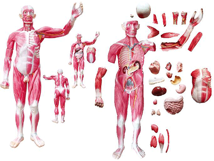
mannequin muscle anatomy
right facial muscles of head and neck
Neck muscles: The neck muscles are divided into superficial neck muscles, hyoid muscles and deep neck muscles according to their location. The sternocleidomastoid muscle is located on both sides of the neck, most of which are covered by the platysma. The function of the sternocleidomastoid muscle is: the contraction of one side of the sternocleidomastoid muscle makes the head flex to the same side, and the face turns to the opposite side; the simultaneous contraction of both sides can make the head tilt back.
back muscles
Superficial dorsal muscle group: The trapezius muscle is triangular on one side, and the two sides merge into a trapezoid. Originating from the external occipital eminence, the nuchal ligament, and all the spinous processes of the thoracic vertebrae, the muscle bundles focus outward and insert at the clavicle, acromion, and spine of the scapula. When contracted, the scapula is brought closer to the spine, the upper muscle bundles contract to lift the scapula (shrug), and the lower muscle bundle contracts to lower the scapula. Trapezius paralysis can cause "collapsed shoulders". The latissimus dorsi originates from the spinous processes of all vertebrae below the 6th thoracic vertebra and the rear of the iliac crest. The muscle bundles concentrate outward and upward, inserting on the lesser tuberosity crest of the humerus. When contracted, the arm adducts, internally rotates and extends backwards, such as Back hands pose.
thoracic abdominal muscles
Pectoralis major, external oblique muscle: The pectoralis major originates from the clavicle, sternum, and the upper 6 costal cartilages. When contracted, the shoulder joint can be adducted, rotated, and flexed. When the upper limbs are immobilized, the torso can be lifted up and assist the inhalation. The external oblique muscle originates from the outside of the lower 8 ribs. The muscle bundle is obliquely forward and downward. The lateral side near the rectus abdominis is transferred to the aponeurosis. White line in the middle of the anterior abdominal wall. The inferior border of the external oblique aponeurosis is thickened and curled, stretching between the anterior superior iliac spine and the pubic tubercle, called the inguinal ligament.
thoracic abdominal muscles
Pectoralis minor, serratus anterior, rectus abdominis, internal oblique, transverse abdominis: The pectoralis minor originates from the 3rd to 5th ribs and inserts into the coracoid process of the scapula. The serratus anterior originates from the 1st to 8th ribs, and the muscle bundle runs backward and upward, inserting on the medial border and lower corner of the scapula. The rectus abdominis originates from the pubic crest and inserts upward at the xiphoid process and the 5th to 7th costal cartilages. The internal oblique muscle bundle spreads out in a fan shape, and transfers to the lateral side of the rectus abdominis as aponeurosis and divides into two layers, wrapping the rectus abdominis, and finally the white line. The transversus abdominis originates from the lower six ribs, the thoracolumbar fascia, the iliac crest and the lateral part of the inguinal ligament. Wire.
Muscles in front of shoulder and upper arm
Anterior brachialis: includes the biceps, coracobrachialis, and brachialis. The biceps muscle is located in the superficial layer of the front of the arm and is fusiform. The origin has two heads, the long head and the short head. The long head originates from the supraglenoid tubercle of the scapula, passes through the shoulder joint capsule, and descends through the intertuberous groove; the short head originates from the coracoid of the scapula. The two ends merge into one muscle belly and descend, inserting on the radial tuberosity. Mainly elbows and shoulders. When the forearm is in pronation, the biceps allow it to be supinated. The coracobrachialis muscle is on the medial side of the biceps muscle, originating from the coracoid process of the scapula and inserting in the middle of the medial side of the humerus. Its function is to flex the shoulder joint. The brachialis is located deep to the lower half of the biceps, originates from the anterior aspect of the lower half of the humeral body, and inserts into the ulnar tuberosity. Its function is to flex the elbow joint.
Back of shoulder and arm muscles
Behind the shoulder muscles and arm muscles: The shoulder muscles are distributed around the shoulder joint, all originating from the upper limb girdle bone and inserting into the humerus. The deltoid muscle is triangular in shape and surrounds the shoulder joint from the anterior, posterior and lateral sides. The supraspinatus muscle arises from the supraspinatus fossa, the teres minor is below the infraspinatus, and the teres major is below the teres minor. The posterior group of the brachial muscles is the triceps capitis, with three heads. The long head originates from the subglalenoid tubercle of the scapula; the lateral and medial heads both originate from the back of the humerus. Synthetic triceps belly, with flat tendon at the ulnar eagle. The main function is to extend the elbow joint, and its long head extends and adducts the shoulder joint.
forearm muscles
The posterior group of the forearm muscles: there are 5 superficial layers, from the radial side to the ulnar side, which are the extensor carpi radialis longus, the extensor carpi radialis brevis, the extensor digitorum, the extensor carpi ulnaris, and the extensor carpi ulnaris. There are also 5 deep layers, from top to bottom, from the radial side to the ulnar side, they are the supinator, abductor pollicis longus, extensor pollicis brevis, extensor pollicis longus, and extensor index finger.
front forearm muscles
Anterior forearm muscle group: There are 9 anterior forearm muscle groups, divided into superficial and deep layers. There are 6 superficial layers, from radial side to ulnar side, they are brachioradialis, pronator teres, flexor carpi radialis, palmaris longus, flexor digitorum superficialis and flexor carpi ulnaris. There are 3 deep layers, namely flexor pollicis longus, flexor deep digitorum, and pronator anterior.
palmar muscles
Hand muscles: Hand muscles are all located on the palm side of the hand and mainly move the fingers. They are divided into lateral, medial and middle groups. The outer group is called the thenar. The medial group is called the hypothenar. The middle group is located between the palm and the metacarpal bones and includes 4 lumbrical muscles and 7 interosseous muscles.
Buttocks and back thigh muscles
Gluteus medius, gluteus minimus, and piriformis: The gluteus medius is located deep to the gluteus maximus, and the gluteus minimus is located deep to the gluteus medius. Both muscles originate outside the ilium wing and insert into the greater trochanter. Both muscles work together to abduct the hip joint. The piriformis muscle originates from the front of the sacrum and exits the pelvis through the greater sciatic foramen into the buttocks, where it inserts at the top of the greater trochanter of the femur. Abduction and external rotation of the hip joint. The foramen magnum is divided by the piriformis muscle into the superior piriformis foramen and the inferior piriformis foramen, through which blood vessels and nerves pass.
Anterior medial thigh muscles
Anterior and medial thigh muscles: The anterior group includes the sartorius and quadriceps. The sartorius muscle originates from the anterior superior iliac spine and inserts on the medial surface of the upper end of the tibia. The quadriceps has four heads called the rectus femoris, vastus medialis, vastus lateralis, and vastus intermedius. The 4 heads merge and move downward as tendons, wrapping around the front and sides of the patella, and then extending downward as the patellar ligament, which inserts into the tibial tuberosity. There are 5 muscles in the medial group, and the superficial layers include the pubis, adductor longus, and gracilis from outside to inside. Deep to the pubic and adductor longus muscles are the short adductor muscles. There is a broad and thick adductor magnus on the deep side.
Muscles of the front inner thigh
Medial group of thigh muscles (deep surface): The medial group all originate from the pubic ramus, ischial ramus, and ischial tuberosity around the obturator foramen. Except for the gracilis, which inserts on the medial side of the upper end of the tibia, all other muscles insert on the thick line of the femur. The adductor magnus also has a tendon that inserts into the adductor tubercle of the medial epicondyle. There is a hole between this tendon and the femur, called the adductor tendon hiatus, through which the large blood vessels of the lower limbs pass. The main function of the medial muscles is to adduction of the thigh.
Muscles of the buttocks and back of the thighs
Gluteus maximus and posterior thigh muscles: The gluteus maximus originates from the outside of the ilium wing and behind the sacrum and inserts into the iliotibial band and the gluteus tuberosity of the femur. The upper 1/4 of the buttocks is the commonly used intramuscular injection site. The long head of the biceps femoris originates from the ischial tuberosity, the short head originates from the femoral thick line, and the two ends merge into the fibular head with a long tendon. The semitendinosus tendon is slender and occupies almost half of the muscle. It originates from the ischial tuberosity along with the long head of the biceps femoris and inserts on the medial side of the upper end of the tibia. The semimembranosus is deep to the semitendinosus and arises from the ischial tuberosity as a membranous flat tendon. The membranous aponeurosis accounts for almost 1/2 of the total length of the muscle, and the lower end inserts on the medial condyle of the tibia.
Anterior lateral calf and dorsal foot muscles
Anterior Calf Muscles: Located in the front of the calf, there are three muscles, from the medial to the lateral, the tibialis anterior, the extensor digitorum longus and the extensor digitorum longus. The tibialis anterior originates from the lateral aspect of the tibia and inserts on the medial cuneiform and the base of the first metatarsal. The extensor digitorum longus originates from the upper end of the fibula and divides downward into four tendons that insert into the middle and distal phalanx of the 2nd to 5th toes, respectively. The extensor longus originates from the anterior aspect of the fibula and the interosseous membrane of the calf, and ends at the base of the distal phalanx of the phalanx. All three anterior calf muscles extend (dorsiflex) the ankle. The extensor digitorum longus and the extensor digitorum longus also extend the 2nd to 5th toes and the extensor digitorum longus, respectively, and the tibialis anterior can also invert the foot.
Anterior lateral calf and dorsal foot muscles
Anterior-lateral group of calf muscles: Located on the outside of the calf, including the peroneus longus and peroneus brevis. The peroneus longus muscle originates from the lateral surface of the fibula 2/3 of the bony surface, and its long tendon goes around the back of the lateral malleolus into the sole of the foot and inserts at the base of the cuneiform and first metatarsal. The peroneus brevis originates from the inferior 2/3 of the lateral surface of the fibula, and the tendon passes behind the lateral malleolus and inserts into the 5th metatarsal tuberosity. The fibula longus and brevis diverters evert the foot and plantarflex the ankle.
muscles behind the calf
The superficial layer of the posterior group of the calf muscles: The superficial layer of the gastrocnemius and soleus muscles is collectively called the triceps calf. The medial and lateral heads of the gastrocnemius originate from the medial and lateral epicondyles of the femur respectively; the deep soleus originates from the back of the upper ends of the tibia and fibula, and the two muscles migrate into the thick Achilles tendon in the middle of the calf and insert into the calcaneal tubercle. The triceps calf plantarflexes the ankle, flexes the calf, and lifts the heel. When standing, the ankle and knee joints can be immobilized to prevent the body from leaning forward.
plantar muscles
Foot muscles: Foot muscles are divided into dorsal and plantar muscles. The dorsal foot muscles include the extensor brevis and extensor digitorum brevis, which help extend the toes. The distribution and function of the plantar muscles are similar to those of the hand muscles, and they can also be divided into medial, lateral, and intermediate groups, but lack the palmar muscles. In addition, there are flexor digitorum short and quadratus plantar muscles, which have the function of flexing the toes and maintaining the arch of the foot.

Eelephone:13506223680 landline:0512-58961302
fax:0512-58961302 Website:www.cnbbmx.com
Address:Bridge No. 4, Leyu Town, Zhangjiagang City, Suzhou City
|
| |
cell phone station | WeChat public account |